 |
Neural Modeling and Biomagnetism
|
|
|
|
Neural Modeling and Biomagnetism
|
|
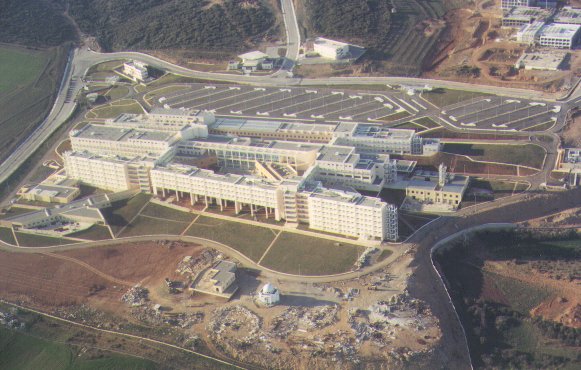
New
University Hospital
|
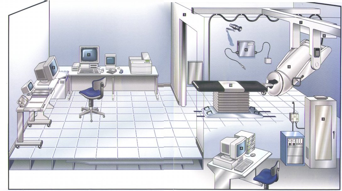
| The
new 122 channel SQUID Click on an image to enlarge |
|
 |
 |
| MEG | EEG |
 |
 |
Photios A. Anninos
Emer Prof. P.Anastasiadis,
Prof. N.Tsagas, Prof. E.Sivridis,
Assistant Prof A. Adamopoulos, Assistant Prof A. Kotini, Dr. P. Tantalakis
Theoretical neural models
to understand the structure and function of the Central
Nervous System (CNS). Some of these models explain memory
functions, topology, abnormal CNS functions, EEG and
models for the electromagnetic radiation from the brain.
Chaos Theory and Epilepsy.
Experimental
Neuroelectrophysiology using intracellular recordings
from the optic center of the cat's brain as well as
Electroencephalographic (EEG) measurements in order to
correlate theoretical and experimental results.
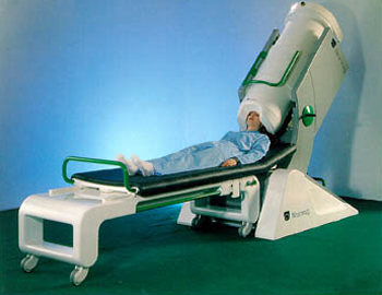
Magnetoencephalographic
(MEG) measurements using SQUID's from patients with CNS
disorders, immune disorders, gynaecological oncology,
breast and ovarian cancer, measurements in perinatal
medicine, normal and preeclamptic pregnancies, and
finally measurements in uremic patients.
The methods employed
in CNS diagnostics are collectively referred to as
"Neuroimaging". The Neuroimaging includes
procedures for investigating both cerebral morphology and
cerebral function.
Such diagnostics are Computed
Tomography (CT), Magnetic Resonance Imaging (MRI),
Positron Emission Tomography (PET) and Single-Photon
Emission Computed Tomography (SPECT).
On the other hand
topographic mapping of electroencephalograms (EEG),
evoked potentials and magnetic fields represent the most
important topographic functional procedures. The latter
applications include not only magnetic fields evoked by
stimuli relating to different sensory modalities, but
also endogenous and motor fields resulting from
spontaneous brain magnetic activity.
Such brain magnetic
activity can be recorded using SQUID technology and it is
called Magnetoencephalograms (MEG) which is the
complement of the EEG. The advantage of recording
electric and magnetic fields over other neuroimaging
procedures is that those techniques are completely
noninvasive and have extremely short analyses times.
In
the list of publications which is followed after this
introduction we would like to demonstrate the importance
of magnetoencephalography (MEG) as diagnostic procedure
used in patients with CNS disorders.
Furthermore some
relevant publications are also listed utilizing the MEG
diagnostic technique in conjuction with the application
of extremely low magnetic fields in order to ameliorate
CNS dysfunctions.
Pineal gland research,
melatonin measurements. Methods of decalcification of
pineal gland.
The use of external magnetic fields to ameliorate CNS disorders.
For more
information or any suggestions you can send your e-mail to: Prof.
P. Anninos or to:George
Charalambidis